The Official OCT Interpretation Optical coherence tomography, Optometry education, Opthalmic

OCT was first introduced in 1991. transparency of the eye (i.e. the viewed through the pupil), OCT invaluable ophthalmic diagnostic tool. to Spectral/Fourier Domain. 3. High scanning. 4. Single axial interference between. 5. Up to 5 microns of.
Anatomy Brisbane Retina Dr Abhishek Sharma
Retinal ultrastructures visualized by OCT. Based upon early TD-OCT, structural correlations with the OCT images were first reported. In 1991, a TD-OCT was capable of producing a single A-scan in 1.25 s with an axial resolution of 17 µm. 25 The inner retina hyper-reflective zone was reported to represent the nerve fibre layer, but the outer retina hypo-reflective zone was not further described.
What is an OCT Scan?
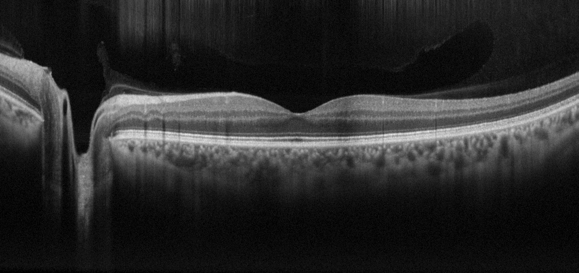
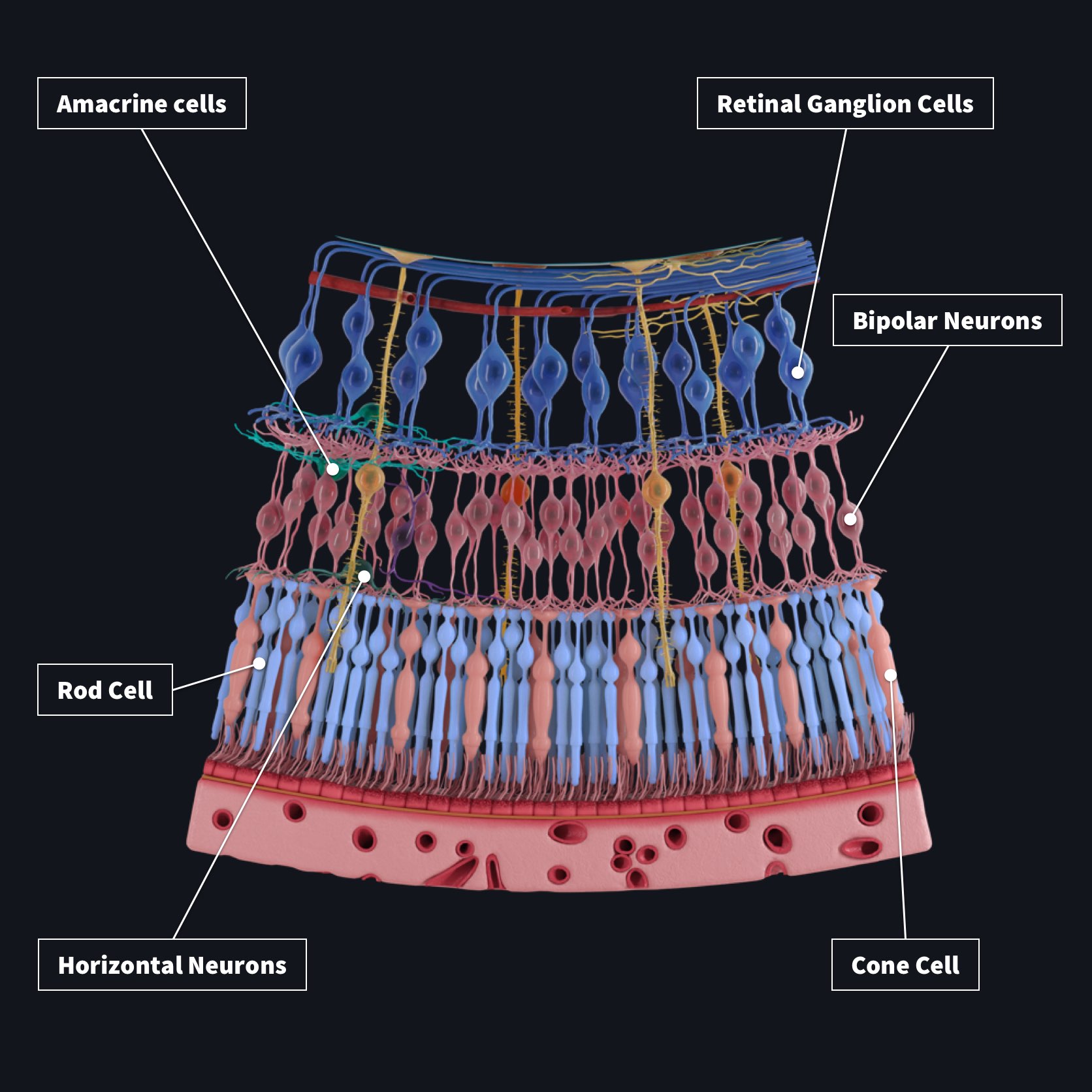
Image courtesy of Discovery Eye Foundation1 Image courtesy of Wikipedia / Public Domain Optical Coherence Tomography (OCT) of the Retina Image courtesy of Christopher M. Putnam2 The retina is a multilaminar structure comprised of separate layers of neurons and glial cells which forms the inner surface of the eye's posterior segment.
Oct Retinal Layers Labeled
Based on this model, we carried out experiments and proposed a new DeepRetina method for the automatic segmentation of retinal layers in retinal OCT images. DeepRetina is based on a deep neural network; we improved Xception 48 and used the improved Xception65 as the backbone network to extract and learn the characteristics of retinal layers.
What Does A Normal OCT Look Like? Eyes On Eyecare

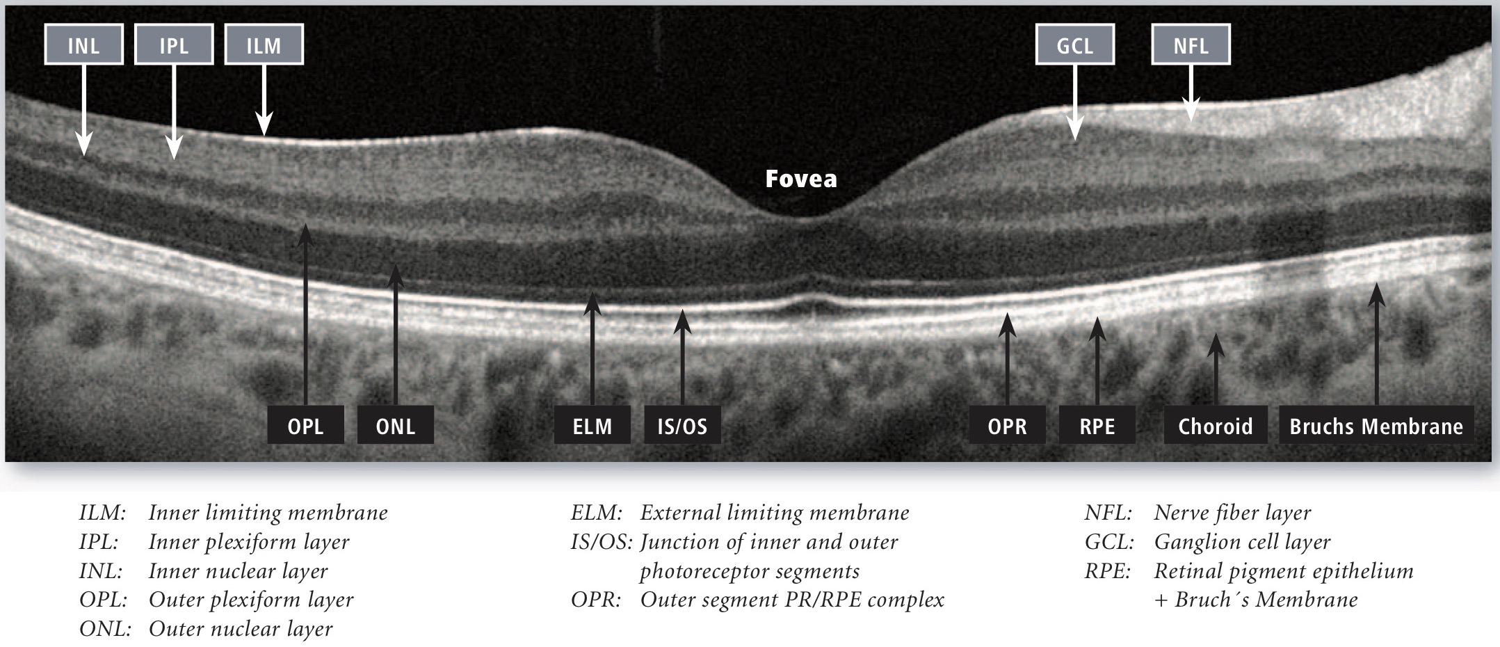
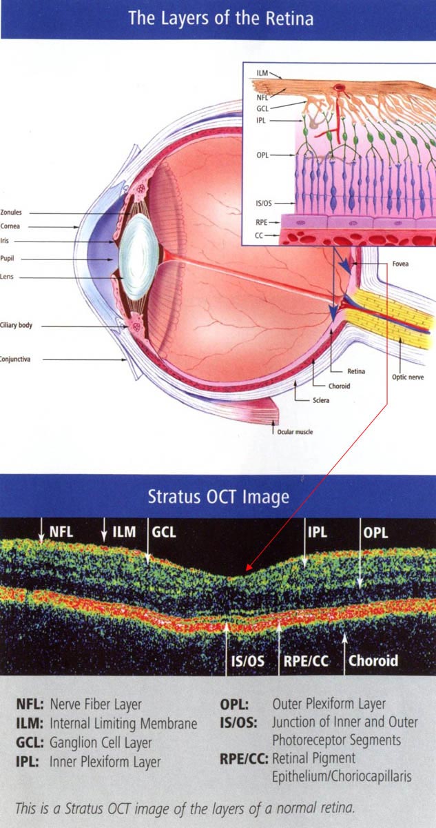
The first step in interpreting optical coherence tomography (OCT) images is understanding the layers of the retina as observed in a "normal" retina, or a retina without macular pathology. The image below is an enlargement of a cross sectional image of a "normal" retina obtained from a spectral domain OCT.
OCT measurements and retinal layers. Visualization of the laminar... Download Scientific Diagram

Normal OCT Anatomy. Istanbul Retina Institute. Advancements in spectral domain optical coherence tomography (SD-OCT) technology enables clear visualization of very small structural details of the posterior segment. Vitreous and vitreoretinal interface: Vitreous (hyporeflective space) Premacular bursa (hyporeflective space in front of the macula)
Normal retina, OCT scan Stock Image C026/7621 Science Photo Library

Optical Coherence Tomography (OCT) is a non-invasive diagnostic technique that renders an in vivo cross-sectional view of the retina. OCT utilizes a concept known as interferometry to create a cross-sectional map of the retina that is accurate to within at least 10-15 microns. OCT was first introduced in 1991 by Huang and colleagues [1] and has.
OCT Illuminating the retina layer by layer Ophthalmology Times
Optical coherence tomography (OCT) is a non-invasive technique for cross-sectional tissue imaging. It typically uses light in the near-infrared spectral range which has a penetration depth of several hundred microns in tissue.. The ability to segment retinal layers allows for thickness measurement, which improves glaucoma diagnosis, because.
(a) Raw OCT image of the retina. (b) Segmented retinal OCT image by... Download Scientific Diagram

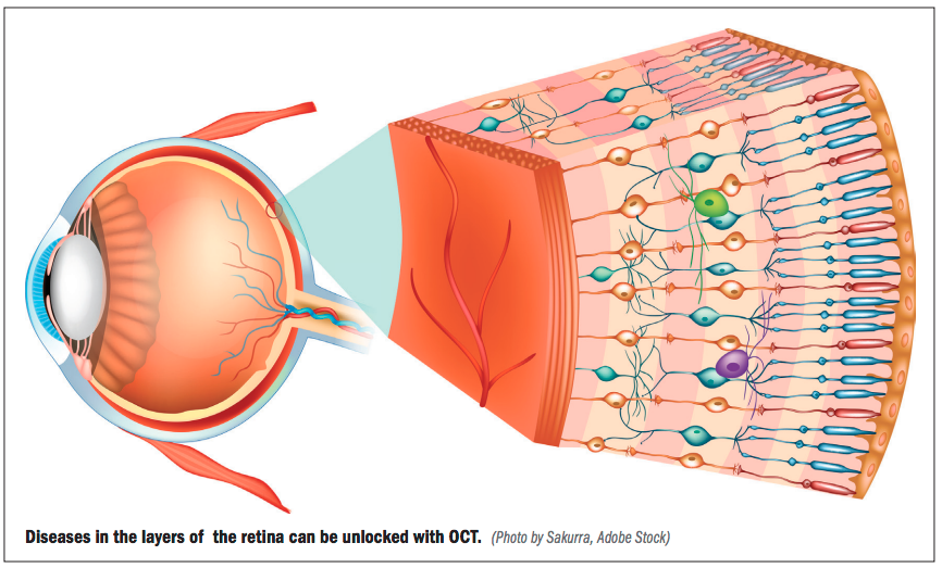
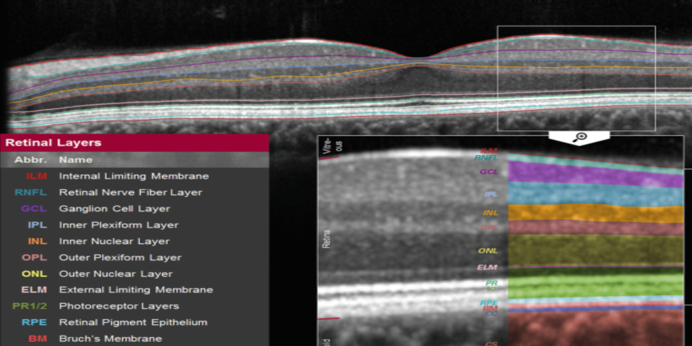
OCT, especially high-resolution spectral-domain (SD-OCT), permits the visualization of all retinal layers, as well as the vitreous and the choroid. Localization of an abnormal finding to a specific layer of the retina often leads to a diagnosis that would not be otherwise possible.
OCT in Ophthalmology Wasatch Photonics
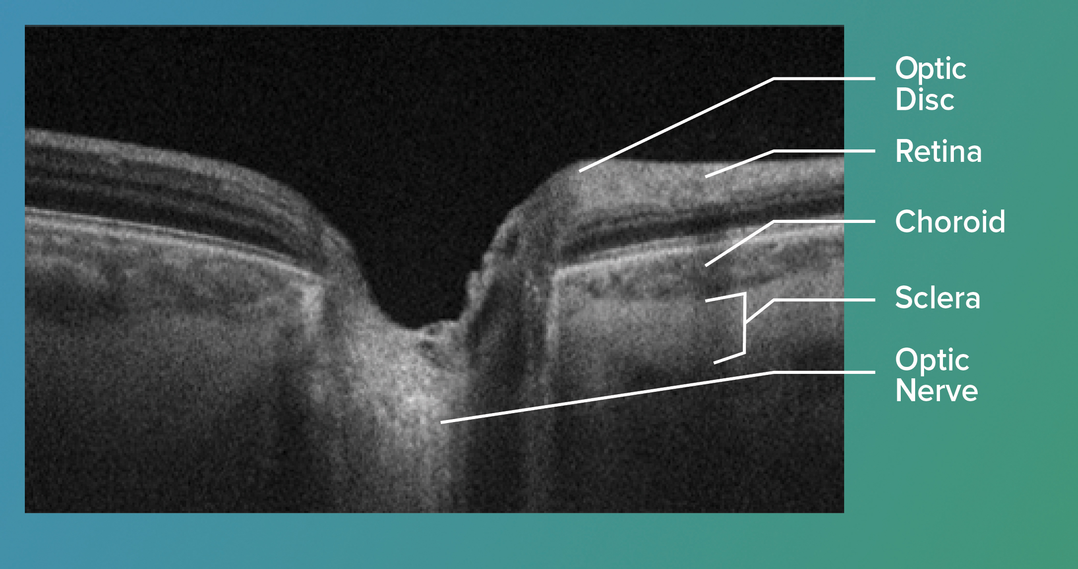
The borders of the choroid-scleral junction can be seen. Posterior segment evaluation with optical coherence tomography (OCT) allows visualization of the vitreous, retinal layers, retinal pigment epithelium (RPE) and the choroidal layers. Figure 1: SD-OCT scan of a normal right eye. The vitreous, retinal layers and choroidal layers are visible.
Optical Coherence Tomography OCT Retina & Optic Nerve Scan South Bay Ophthalmology
Optical Coherence Tomography. Optical coherence tomography (OCT) is a noninvasive imaging method that uses reflected light to create pictures of the back of your eye. It can be used to diagnose and manage diseases like diabetes-related retinopathy and glaucoma. Contents Overview Test Details Results and Follow-Up.
Segmentation of retinal layers in OCT images with graph theory YouTube

Optical coherence tomography angiography (OCTA) can image the retinal vasculature in vivo, without the need for contrast dye. This technology has been commercially available since 2014, however, much of its use has been limited to the research setting. Over time, more clinical practices have adopted OCTA imaging. While countless publications detail OCTA's use for the study of retinal.
OCT Image Analysis
Optical coherence tomography (OCT) is an optical analog of ultrasound imaging that uses low coherence interferometry to produce cross-sectional images of the retina. It captures optical scattering from the tissue to decode spatial details of tissue microstructures. It uses infrared light from a super-luminescent diode that is divided into two.
Layers Of Retina On Oct

1. Margolis R, Spaide RF. A pilot study of enhanced depth imaging optical coherence tomography of the choroid in normal eyes. Am J Ophthalmol. 2009;147(5):811-5. 2. Zhou Y, Song M, Zhou M, et al. Choroidal and retinal thickness of highly myopic eyes with early stage of myopic chorioretinopathy: Tessellation. J Ophthalmol. 2018;2181602. 3.
Optical Coherence Tomography

The OCT-A is capable of separately evaluating a layer of any thickness and depth (slabs) in the retina and choroid, making it especially useful in the study of the capillary layers of the retina. Unlike conventional "2-D" angiograms, OCT-A technology makes it possible to obtain "3-D" images of the macula and visualize the peripapillary.
SS OCT with the horizontal line examination shows preserved retinal... Download Scientific Diagram

I maging with optical coherence tomography (OCT) has become standard in retina care. Its widespread use can be attributed to its ability to deliver a vast amount of high-resolution information regarding the structure of the retina both quickly and safely. A strength of OCT is its ability to segment various retina layers, which enables cross.
.

